Digital Mammography & Tomosynthesis (3D Mammography)
The mammography is the primary preventive imaging examination for the early diagnosis of breast cancer. It is the only method that has been proven to contribute to the reduction of mortality from the disease, as it can detect malignancies 1–4 years before they become clinically apparent.
What is digital mammography?
The digital mammography uses low-dose radiation to produce high-resolution images of the breast. Advances from 1965 to the present have significantly improved image quality while simultaneously reducing radiation dose. It remains the first-line examination for population-based preventive screening, but it has certain limitations, such as a small probability of failing to detect some types of cancer (1 in 5).
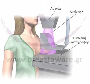
How is mammography performed?
The procedure is simple and lasts about 20 minutes. Each breast is compressed for a few seconds, and two projections (frontal and lateral). In some cases, additional more images (magnification or spot compression), for better analysis of areas of interest. The radiology report is usually available within 7–10 days.
How much radiation do I receive?
The radiation emitted during digital mammography is extremely low, equivalent to natural background radiation absorbed over three months or after a two-hour flight. Scientific data do not link mammography to increased cancer risk, even with annual screening from age 40 onward.
What should I keep in mind before the exam?
- Ideal interval: 7th to 15th day of the cycle, for less sensitivity.
- Do not use deodorants or antiperspirants on the day of the exam.
- Choose specialized radiologist center with experience in mammography.
- Always provide the previous exams for comparison.
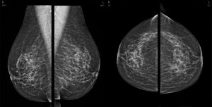
What are suspicious mammographic findings?
The Breast imaging can reveal:
- Masses
- Microcalcifications
- Asymmetries
- Architectural distortions
These findings may require monitoring or additional control.
What is Digital Tomosynthesis (3D Mammography)?
The breast tomosynthesis is an advanced form of digital mammography that offers 3D visualization of the breast. The X-rays move in an arc and create multiple images, which are processed to give clearer and more complete picture of tissues.
Large studies show that tomosynthesis:
- Increases breast cancer detection, especially in women with dense breasts,
- Reduces false positives and the number of reassessments,
- Έχει be included in population screening programs in many countries.
Although radiation dose is slightly higher than conventional mammography, it remains within safe limits. Tomosynthesis is increasingly recommended as the ideal option for women over 40,with dense breasts or or moderate to high risk of breast cancer.
What if I am pregnant?
If there is a possibility of pregnancy, inform the radiology department. Although the radiation dose is low, screening is usually is postponed for women without increased risk.
What about after surgery or implants?
- After lumpectomy, mammography is performed 6–12 months post-radiotherapy.
- After mastectomy, no imaging is done on the operated side.
- If there is plastic restoration or silicone implant, special images are taken to clearly visualize the gland.
Conclusion
The digital mammography and tomosynthesis (3D mammography) are valuable tools in the breast cancer screening. The choice of the appropriate test depends on the age, breast density, and individual history of each woman.
Learn more at breastaware.gr
At breastaware.gr, you will find reliable, scientifically validated, and easy-to-understand information on all aspects of breast health — from prevention to diagnosis and follow-up. Our goal is to empower every woman to care for herself with knowledge, consistency, and confidence.
Bibliography:
- Friedewald SM et al. Breast cancer screening using tomosynthesis in combination with digital mammography, JAMA 2014
- Bernardi D et al. Breast cancer screening with tomosynthesis (3D mammography) with acquired or synthetic 2D mammography compared with 2D mammography alone (STORM-2): a population-based prospective study The Lancet Oncology 2016
- Conant EF et al. Five Consecutive Years of Screening with Digital Breast Tomosynthesis: Outcomes by Screening Year and Round, Radiology 2020
- Gilbert FJ et al. Accuracy of Digital Breast Tomosynthesis for Depicting Breast Cancer Subgroups in a UK Retrospective Reading Study (TOMMY Trial), Radiology 2015
- Philpotts LE et al. Advanced 3D Mammography Detects More Breast Cancers, Fewer False Positives, Radiology 2024