Palpable Breast Mass: When Should I Be Concerned?
The discovery of apalpable mass in the breastis an event that often causes concern. Whether it is detected by the woman herself duringbreast self-palpation, or arises during preventive screening or by a physician, it raises reasonable questions: Is it something serious? Could it beBreast cancer;
The reassuring news is that most palpable breast masses arebenign. However, proper and timely investigation is essential — not to cause alarm, but to ensure accurate knowledge.
What is a palpable mass?
A palpable mass is a lump or nodule distinct from the normal breast tissue, often characterized by different firmness or asymmetry. It may be felt on touch or accompanied by other symptoms, such as:
- Pain or tenderness
- Nipple discharge (bloody, white, or green)
- Redness or skin changes over the breast
- Nipple retraction
When should I seek evaluation?
Even if the mass is painless, it should not be ignored. During medical assessment, the physician will consider:
- Duration of the mass
- Whether it varies with the menstrual cycle
- Any changes in breast appearance
- Personal or family history of breast cancer

What diagnostic tests are necessary for a mass?
Diagnosis combinesclinical breast examination with imaging assessmentand, if necessary, biopsy.
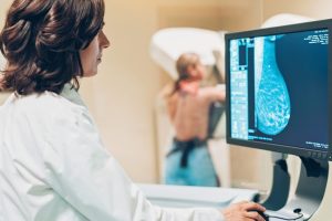
- Digital mammography
The first-line imaging for women over 35 years. Detects suspicious features associated withbreast cancer, even in asymptomatic cases.
- Breast ultrasound:
Preferred in younger women (<30 years), pregnancy, or lactation due to dense breast tissue. Differentiates cystic, solid, or mixed masses.
- Breast MRI:
Used selectively when other imaging is inconclusive. Offers higher sensitivity but increased false positives.

Definitive diagnosis: The role of biopsy
No imaging modality can replace biopsy when there is suspicion ofmalignancy. The options include:
- Fine needle aspiration (FNA) or core needle biopsy, often ultrasound-guided
- Surgical biopsy (in selected cases)
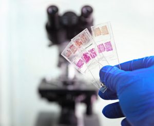
Histopathological examination determines whether the lesion is benign (e.g., fibroadenoma, cyst, fat necrosis) or malignant, guiding appropriate treatment or surveillance.
Conclusion: Don’t ignore the signs — Act responsibly
Your body gives you signals. Apalpable lump in the breastdoes not necessarily meancancer, but it should always be properly evaluated. The earlier a potential problem is detected, the greater the range of safe and effective treatment options.
Why get informed at breastaware.gr?
Atbreastaware.gryou will find reliable and comprehensible information on breast cancer prevention, diagnosis, and care. We are here to help you care for yourself with knowledge and confidence.
Bibliography:
- American Cancer Society. Breast Lumps. 2023.
- O’Flynn EA, Wilson AR, Michell MJ. Image-guided breast biopsy: state-of-the-art. Clin Radiol. 2010;65(4):259–70.
- Berg WA, Blume JD, Cormack JB, et al. Combined screening with ultrasound and mammography vs mammography alone in women at elevated risk of breast cancer. JAMA. 2008;299(18):2151–63.
- Elmore JG, Armstrong K, Lehman CD, Fletcher SW. Screening for breast cancer. JAMA. 2005;293(10):1245–56.
- Saslow D, Boetes C, Burke W, et al. American Cancer Society guidelines for breast screening with MRI as an adjunct to mammography. CA Cancer J Clin. 2007;57(2):75–89.
